RIDITM: Rapid Infectious Disease Identification by Sequencing

Pan-Bacterial (Bacteria/Archaea) DNA Analysis AND Pan-Eukaryotic (Protozoa/Fungi) DNA Analysis

One test to detect all known bacteria, archaea, and, pathogenic fungi and protozoans.

Our revolutionary RIDITM software screens many different sample types for the presence of all known bacteria, archaea, pathogenic fungi, and protozoans. Utilizing our proprietary Next Generation Sequencing chemistry and bioinformatics analysis, our RIDITM software is capable of identifying organisms at the species level or the nearest known relative. This test requires that the organisms be present in the sample provided and does not discriminate between viable or dead organisms. Potential novel organisms are flagged and the nearest known relative is identified. RIDITM is more sensitive and accurate than standard single/multi-organism PCR panels and fickle microbial culturing, reducing the risk of false positives, false negatives, and speculation over appropriate treatment. By identifying the specific organisms, RIDITM provides the critical data patients and providers need to make informed health decisions.

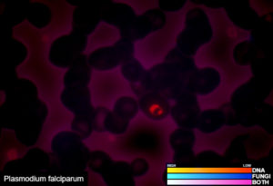
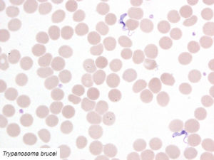
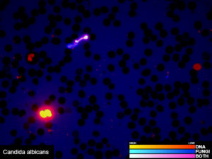
Examples of validated organisms include, but are not limited to: Staphylococcus, Streptococcus, Bartonella, Borrelia, Trypanosoma, Giardia, Acanthamoeba, Prototheca, Leishmania, Babesia, Cryptococcus, Cryptosporidium, Blastocystis, Entamoeba, etc. For more information about DNA sequencing, click here.

Order Test Kit

Microscopy Tests

Mosaic Stain (Fluorescent Fungal And DNA Stains):

This assay is only performed by Fry Laboratories, LLC. Two fluorescent stains are used to simultaneously highlight DNA positive material in addition to potential fungal structures in a wet mount blood preparation. This allows for more effective fungal contribution assessment while simultaneously screening for other non-fungal organisms. Simply, DNA positive structures are labeled by a single color stain indicating non-fungal stuctures (shown in red). Structures containing fungal material are labeled with a blue dye. Both image channels are captured and a combined photograph and subsequent report are generated based upon the observations. The combined image shows DNA positive material in a red color and fungal material in a blue color; with overlapping patterns in a magenta color.

Advanced Stain (Fluorescent DNA Stain):

This test is only available from Fry Laboratories, L.L.C. By using a highly specific fluorescent DNA dye, infections and blood-borne biofilm communities can be visualized. This screening test is used to identify structures that stain positive for DNA. We add our dye directly to a sample of blood and observe it under a fluorescent microscope where a photograph and report are then generated based upon the technologist’s findings. Additionally, if no abnormal results are found from the advanced stain procedure, an automatic reflex enrichment test (Organismal Enrichment) is performed to absolutely ensure we can detect any abnormalities. This protocol is to produce a higher yield of detectable infections and biofilm structures.

Giemsa & Modified May-Grünwald Stain:

These two smears are a general screening test used to detect blood-borne infections. The Modified May-Grünwald (MMG) stain was developed by Fry Laboratories to assist in the detection blood-borne infections. The stain is a combination of two unique stains to visualize red blood cells and any other foreign bodies. While the Giemsa staining procedure is a test that has long standing industry standard used to analyze blood with both a thick and thin blood preparation. The thin prep involves dragging a spot of blood across a slide using a second slide. The thick prep is simply a drop of blood that is ground into the slide and allowed to dry with no smearing. Both techniques are used in conjunction to determine if there are any abnormal findings in clinical sample. A photograph and report are generated based upon the technologist’s findings.

Order Test Kit
Microscopy Tests at Fry Laboratories LLC

Lyme Line Blot Serology

Serology testing is performed to examine a patient’s immune response (antibodies) to a specific pathogen. Disease causing organisms usually display antigens (foreign molecules presented on their surface) that are detected by the host’s immune system. Both IgG and IgM antibodies in an immunocompetent individual (typical normal immune response) will be produced at specific time points throughout the course of infection. IgM antibodies are the first type to respond to an infection. Their production peaks around the first week after initial infection. IgG antibodies peak between 3-4 weeks post infection. IgG antibodies usually become detectable within 3 weeks following infection and peaks between 2-6 months. Individuals being treated with antibiotics may not develop antibody titers or will develop low antibody levels. It is suggested that patients be off antibiotics for two weeks prior to testing; however, this is subject to clinical necessity. The ability to detect both types of antibodies allows a physician to determine the time course of an infection and whether said infection is active or not.

Serology Tests at Fry Laboratories LLC

Lyme Line Blot IgG And IgM:

This test detects antibodies to various proteins to the Lyme spirochete – Borrelia burgdorferi. Borrelia proteins (also known as antigens) are attached to a thin strip of material (blot) and the patient’s serum containing antibodies are allowed to bind to the proteins. We test for both IgG and IgM antibodies and follow the Center for Disease control criteria for Lyme disease. A positive test is indicated with two or more IgM bands or 5 or more IgG protein bands. It has been reported that patients with Lyme disease produce antibodies of the IgM class during the first weeks after onset of EM (rash) but produce IgG antibodies more slowly. Strips that have 5 (or more) out of 10 significant bands for IgG and 2 out of 3 significant bands for IgM are considered positive for antibodies to B. burgdorferi. Individuals being treated with antibiotics may not develop titers or will develop low antibody levels. It is suggested that patients be off antibiotics for two weeks prior to testing; however, this is subject to clinical necessity. For more information about Lyme Disease and the organism that causes it, for the CDC’s Website click here.